
A standard identity test on a certificate of analysis compares the measured mass of a peptide with the mass expected from its sequence. That is a strong check, but it has a blind spot: two peptides with the same amino acids in a different order have exactly the same mass. Tandem mass spectrometry, written MS/MS, closes most of that gap by breaking the peptide into fragments and reading the sequence from them. This guide explains how it works and what it still cannot see.
From one mass to a ladder of fragments
In MS/MS, the instrument first selects the intact peptide ion, then breaks it apart and measures the masses of the pieces. The resulting fragment spectrum is what gets interpreted, either by matching it against expected fragments or by working the sequence out directly, an approach called de novo sequencing [1].
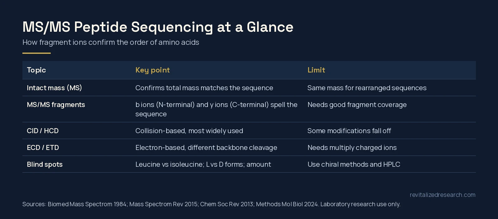
The fragments follow a pattern. When the peptide backbone breaks, the piece that keeps the N-terminus and the piece that keeps the C-terminus are given standard letters. The common nomenclature for these sequence ions was proposed in 1984 and is still used today [2]. In collision-based fragmentation, the main series are called b ions (N-terminal pieces) and y ions (C-terminal pieces). The mass difference between neighbouring ions in a series matches one amino acid, so a complete series spells out the sequence residue by residue.
Ways of breaking a peptide
How the peptide is fragmented changes what you see. Collision-induced dissociation (CID), where the peptide ion is energized by collisions with a neutral gas, is the most commonly used MS/MS activation method [3].
Electron-based methods work differently. In electron capture and electron transfer dissociation (ECD and ETD), multiply charged peptide ions take up an electron, either a free low-energy electron or one transferred from a reagent anion, and the backbone breaks along different pathways [4]. ETD was demonstrated in a linear ion trap in 2004 by transferring electrons from anthracene anions to multiply protonated peptides [5]. These alternative methods, along with higher-energy collision dissociation, have improved identification of certain classes of peptides and modifications, and modern instruments can alternate between fragmentation modes automatically [6].
| Method | How it fragments | Typical strength |
|---|---|---|
| CID | Collisions with a neutral gas | Most widely used; well-understood b and y ion series |
| HCD | Higher-energy collisions | Clear fragment spectra on high-resolution instruments |
| ECD / ETD | Electron capture or transfer to a multiply charged ion | Different backbone cleavage; helpful for some modifications and larger peptides |
Reading the sequence: database match or de novo
For a synthetic peptide, the expected sequence is already known, so the fragment spectrum can be checked directly against the fragments that sequence should produce. De novo sequencing goes further and derives the sequence from the spectrum without a template. It has a long history: in 1999 one group described an algorithm for automated de novo interpretation, noting that manual interpretation by experts had been the norm [7]. Precision instruments changed the picture; peptide de novo sequencing performed dramatically better with high-resolution mass data than with low-precision ion-trap data [8].
Experts still emphasize understanding the spectra. A tutorial on de novo sequencing from CID data points out that many known rules of peptide fragmentation are not built into search engine scoring, so familiarity with the raw data helps a researcher evaluate results critically [3]. De novo methods are also useful when modifications are present that a simple sequence match would miss [9].
What MS/MS still cannot tell apart
- Leucine and isoleucine. These two amino acids have identical masses, so standard fragmentation cannot distinguish them from mass alone.
- L and D forms. A residue flipped to its mirror image has the same mass and usually the same fragments. Chiral purity needs dedicated methods, as described in our guide to peptide racemization and chiral purity.
- How much of each component. MS/MS identifies; it is not a purity measurement. Purity still comes from HPLC, as covered in our HPLC vs mass spectrometry guide.
Where it fits on a COA
Most routine identity testing for short synthetic peptides relies on an intact mass match, which is fast and usually sufficient. MS/MS adds value when a sequence has a same-mass alternative, when an impurity needs to be identified, or when the position of a modification matters, such as the end groups covered in our guide to C-terminal amidation and N-terminal acetylation. To see what Revitalized Research reports for each batch, browse the COA library or our guide on how to read a peptide COA.
All Revitalized Research products are sold for laboratory research use only. They are not for human or veterinary use. This article describes analytical chemistry and does not make any claim about biological effects.
Frequently asked questions
What is the difference between MS and MS/MS for peptides?
A single-stage mass measurement gives the mass of the intact peptide. MS/MS selects that peptide, breaks it into fragments and measures them, which lets the analyst confirm the order of the amino acids, not just the total mass.
What are b and y ions?
They are the two main fragment series produced when a peptide backbone breaks in collision-based MS/MS. b ions keep the N-terminus and y ions keep the C-terminus, and the mass steps between neighbouring ions correspond to individual amino acids.
What is the difference between CID and ETD?
CID breaks peptide ions by collisions with a neutral gas and is the most common method. ETD transfers an electron to a multiply charged peptide ion, which breaks the backbone along different pathways and can help with some modifications and larger peptides.
What is de novo peptide sequencing?
It is working out a peptide’s sequence directly from its fragment spectrum, without matching it to a known sequence or database. High-resolution instruments have made it much more reliable.
Can MS/MS tell leucine from isoleucine?
Not with standard fragmentation, because the two amino acids have the same mass. Distinguishing them needs specialized fragmentation approaches or other analytical methods.
References
- Hjernø K, Højrup P (2024). Interpretation of Tandem Mass Spectrometry (MS-MS) Spectra for Peptide Analysis. Methods Mol Biol. PMID: 38997483. doi: 10.1007/978-1-0716-3914-6_8.
- Roepstorff P, Fohlman J (1984). Proposal for a common nomenclature for sequence ions in mass spectra of peptides. Biomed Mass Spectrom. PMID: 6525415. doi: 10.1002/bms.1200111109.
- Medzihradszky KF, Chalkley RJ (2015). Lessons in de novo peptide sequencing by tandem mass spectrometry. Mass Spectrom Rev. PMID: 25667941. doi: 10.1002/mas.21406.
- Zhurov KO, Fornelli L, Wodrich MD et al. (2013). Principles of electron capture and transfer dissociation mass spectrometry applied to peptide and protein structure analysis. Chem Soc Rev. PMID: 23450212. doi: 10.1039/c3cs35477f.
- Syka JE, Coon JJ, Schroeder MJ et al. (2004). Peptide and protein sequence analysis by electron transfer dissociation mass spectrometry. Proc Natl Acad Sci U S A. PMID: 15210983. doi: 10.1073/pnas.0402700101.
- Guthals A, Bandeira N (2012). Peptide identification by tandem mass spectrometry with alternate fragmentation modes. Mol Cell Proteomics. PMID: 22595789. doi: 10.1074/mcp.R112.018556.
- Dancík V, Addona TA, Clauser KR et al. (1999). De novo peptide sequencing via tandem mass spectrometry. J Comput Biol. PMID: 10582570. doi: 10.1089/106652799318300.
- Frank AM, Savitski MM, Nielsen ML et al. (2007). De novo peptide sequencing and identification with precision mass spectrometry. J Proteome Res. PMID: 17203955. doi: 10.1021/pr060271u.
- Standing KG (2003). Peptide and protein de novo sequencing by mass spectrometry. Curr Opin Struct Biol. PMID: 14568614. doi: 10.1016/j.sbi.2003.09.005.