
Two research peptides can leave the synthesis line with the same sequence and the same purity, then look different on a chromatogram months later. In most cases the reason is chemical: some molecules in the vial have changed. The two changes that show up most often are oxidation and deamidation. They affect different amino acids, they shift the molecular mass by very different amounts, and they need different analytical tools to find. This guide explains both, side by side, so a certificate of analysis (COA) is easier to read.
Degradation products like these are one of the main families of peptide-related impurities, next to synthesis by-products such as deletion and insertion sequences [1]. Both pathways are covered in long-standing stability literature for peptides and proteins, in solution and in the dried state [2].
Oxidation: what changes and where
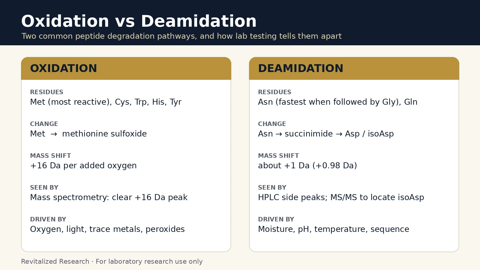
Oxidation adds oxygen to reactive side chains. Methionine, cysteine, histidine, tryptophan and tyrosine are the residues most susceptible, because they react readily with reactive oxygen species [3]. The same review lists the usual triggers: contaminating oxidants (for example peroxides), transition metal ions that catalyze the reaction, and light. Oxidation can also be influenced by pH, temperature and buffer composition [3].
For methionine, the main product is methionine sulfoxide. Each added oxygen raises the molecular mass by 16 Da, which makes oxidation one of the easier changes to spot by mass spectrometry. In a light-exposure study, the oxidized material showed mass increases of 32 Da (two methionines oxidized), while chemically oxidized material showed increases of 16 Da [4].
One caution for anyone reading a mass spectrum: the analysis itself can oxidize methionine. Sample preparation and ionization can create a small sulfoxide signal that was not in the vial, which is why dedicated methods exist to measure true oxidation levels in synthetic peptides [5]. A minor +16 Da signal is a reason to look at the method and the HPLC trace together, not proof by itself that a lot has degraded.
Deamidation: what changes and where
Deamidation removes the amide group from asparagine (Asn) and, more slowly, from glutamine (Gln). For asparagine, the backbone nitrogen attacks the side chain and forms a five-membered succinimide ring. The ring then opens to give normal aspartate (Asp) or its isomer, isoaspartate (isoAsp). In a classic model peptide, Val-Tyr-Pro-Asn-Gly-Ala, this happened with a half-life of only 1.4 days at 37 °C and pH 7.4 [6].
Sequence matters a great deal. In a series of model peptides, asparaginyl residues formed succinimides 13 to 36 times faster than aspartyl residues, and a glycine on the following position made the reaction 6.5 to 17.6 times faster than an alanine [7]. Rates for whole proteins can be estimated from the sequence and the local three-dimensional environment of each asparagine [8]. Aspartate can also isomerize through the same succinimide, so a peptide with no asparagine at all can still form isoaspartate [9].
Detection is the hard part. Deamidation raises the mass by just under 1 Da (+0.98 Da), and isoaspartate has exactly the same mass as aspartate. The products are a mixture of succinimide, aspartyl and isoaspartyl forms, with L-isoaspartate usually dominating [9]. A 2026 study found that conventional chromatography and standard mass spectrometry often fail to separate these nearly identical species, and showed that tandem mass spectrometry (CID and ETD fragmentation) can tell them apart. It also confirmed isoaspartate formation under mildly basic conditions such as phosphate-buffered solutions [10].
Oxidation vs deamidation at a glance
| Oxidation | Deamidation | |
|---|---|---|
| Residues | Met (most common), Cys, His, Trp, Tyr | Asn (fastest before Gly), Gln; Asp can isomerize too |
| Product | Methionine sulfoxide and other oxidized forms | Succinimide, then Asp or isoAsp |
| Mass change | +16 Da per oxygen | About +0.98 Da; isoAsp vs Asp is 0 Da |
| Best detected by | Mass spectrometry, confirmed against the HPLC trace | HPLC side peaks, then tandem MS to place the site |
| Main drivers | Oxygen, peroxides, metal ions, light | Moisture, pH, temperature, neighboring residue |
| Artifact risk | Can be created during sample preparation | Can continue in buffered solutions during analysis |
Reading a sequence for these sites
The sequence printed on a product page tells you where these reactions could happen. It does not tell you whether they have happened in a given lot; only that lot’s test data can do that. A few examples from our own catalog:
- Semax (Met-Glu-His-Phe-Pro-Gly-Pro) starts with a methionine, the residue most prone to oxidation, and also carries a histidine.
- MOTS-c (MRWQEMGYIFYPRKLR) contains two methionines, a tryptophan and two tyrosines, plus one glutamine.
- TB-500 contains a methionine, an asparagine, several glutamines and several aspartates, so both pathways are relevant.
- BPC-157 (GEPPPGKPADDAGLV) has no methionine or asparagine, but its two adjacent aspartates are candidates for isomerization.
- Selank (Thr-Lys-Pro-Arg-Pro-Gly-Pro) contains none of the residues discussed here.
Lyophilized does not mean frozen in time
Freeze-drying slows these reactions a lot, but the dried solid is still chemically active. In a study of a lyophilized aspartyl model peptide, residual moisture, temperature and especially the type of bulking agent had significant effects on how fast the peptide degraded in the solid state [11]. Stability in the dried state is discussed as its own topic in the stability literature for this reason [2].
For lab handling, this points to the familiar basics covered in our peptide storage basics guide: keep vials sealed and dry, store them cold and away from light, limit how often a vial is opened, and record when each lot was received.
What to look for on a certificate of analysis
- A chromatogram, not only a number. Oxidized and deamidated species often sit as small peaks next to the main peak. A trace lets you see them; a single purity figure does not.
- An observed mass next to the expected mass. A clean match supports identity. A visible +16 Da companion suggests oxidation; a shift of about 1 Da is easy to miss at low resolution.
- The test date. Both reactions continue over time, so a recent report says more about the material you receive.
- A matching lot number. The data only describes the batch it was run on.
For more on how purity and identity methods differ, see HPLC vs mass spectrometry, and for checking that a report belongs to your lot, see how to verify a peptide COA. Batch reports for our catalog are in the Revitalized Research COA library.
All Revitalized Research products are sold for laboratory research use only. They are not for human or veterinary use. This article describes analytical chemistry and does not make any claim about biological effects.
Frequently asked questions
What is the difference between peptide oxidation and deamidation?
Oxidation adds oxygen to reactive side chains, most often methionine, and raises the mass by 16 Da per oxygen. Deamidation converts asparagine (and more slowly glutamine) into aspartate or isoaspartate through a succinimide intermediate, which raises the mass by only about 1 Da.
Which amino acids are most likely to oxidize?
Methionine, cysteine, histidine, tryptophan and tyrosine are the residues most often cited as susceptible to oxidation, with methionine the most common site in peptides that contain it.
Why is deamidation harder to detect than oxidation?
The mass change is under 1 Da, and isoaspartate has exactly the same mass as aspartate. Standard HPLC and single-stage mass spectrometry can miss these species, so tandem mass spectrometry is used when the exact site has to be identified.
Can a lyophilized peptide still degrade?
Yes. Studies of freeze-dried model peptides show that residual moisture, temperature and the type of bulking agent all change how fast aspartyl and asparaginyl residues react in the solid state. Dry, cold and sealed storage slows these reactions but does not stop them.
Does a methionine in the sequence mean a batch is oxidized?
No. A susceptible residue only tells you where a change could occur. Whether a specific lot contains oxidized material is answered by that lot’s HPLC chromatogram and mass spectrum on its certificate of analysis.
References
- D’Hondt M, Bracke N, Taevernier L et al. (2014). Related impurities in peptide medicines. J Pharm Biomed Anal. PMID: 25044089. doi: 10.1016/j.jpba.2014.06.012.
- Manning MC, Chou DK, Murphy BM et al. (2010). Stability of protein pharmaceuticals: an update. Pharm Res. PMID: 20143256. doi: 10.1007/s11095-009-0045-6.
- Li S, Schöneich C, Borchardt RT (1995). Chemical instability of protein pharmaceuticals: Mechanisms of oxidation and strategies for stabilization. Biotechnol Bioeng. PMID: 18623513. doi: 10.1002/bit.260480511.
- Liu H, Gaza-Bulseco G, Zhou L (2009). Mass spectrometry analysis of photo-induced methionine oxidation of a recombinant human monoclonal antibody. J Am Soc Mass Spectrom. PMID: 19103498. doi: 10.1016/j.jasms.2008.11.011.
- Hoare M, Tan R, Welle KA et al. (2024). Methionine Alkylation as an Approach to Quantify Methionine Oxidation Using Mass Spectrometry. J Am Soc Mass Spectrom. PMID: 38324783. doi: 10.1021/jasms.3c00337.
- Geiger T, Clarke S (1987). Deamidation, isomerization, and racemization at asparaginyl and aspartyl residues in peptides. Succinimide-linked reactions that contribute to protein degradation. J Biol Chem. PMID: 3805008.
- Stephenson RC, Clarke S (1989). Succinimide formation from aspartyl and asparaginyl peptides as a model for the spontaneous degradation of proteins. J Biol Chem. PMID: 2703484.
- Robinson NE, Robinson AB (2001). Prediction of protein deamidation rates from primary and three-dimensional structure. Proc Natl Acad Sci U S A. PMID: 11296285. doi: 10.1073/pnas.071066498.
- Yang H, Zubarev RA (2010). Mass spectrometric analysis of asparagine deamidation and aspartate isomerization in polypeptides. Electrophoresis. PMID: 20446295. doi: 10.1002/elps.201000027.
- Erckes V, Rendueles LC, Misiek A et al. (2026). Revealing deamidation and isoaspartate formation during peptide analysis, purification and storage by tandem mass spectrometry. RSC Med Chem. PMID: 41541711. doi: 10.1039/d5md01025j.
- Oliyai C, Patel JP, Carr L et al. (1994). Chemical pathways of peptide degradation. VII. Solid state chemical instability of an aspartyl residue in a model hexapeptide. Pharm Res. PMID: 7937533. doi: 10.1023/a:1018998312503.